


If you already have dental X-rays and want to know what else is needed before an implant, Blanc Dental Center at 5001 Rue Jean-Talon O in Montréal is a practical place to start. Book a consultation for the specific implant question, bring your existing images, and tell us when and why they were taken. Our dentists can review those images alongside an examination and explain whether further imaging, assessment of your gums, or other treatment is needed to plan the replacement tooth.
You do not need to decide for yourself whether an old panoramic X-ray, a small dental X-ray, or a previous 3D scan is enough. That decision depends on what the image shows, the condition of the implant site, and the restoration you hope to receive. Our Jean-Talon clinic has CBCT 3D imaging and intraoral scanning available for implant assessment when the clinical findings call for them. A consultation does not automatically mean every image must be retaken.
To arrange a visit at 5001 Rue Jean-Talon O, call (514) 564-4554 or use our appointment page. Say that you have existing dental X-rays and want to understand whether they are sufficient for implant planning. The clinic has free parking.
The important distinction: An X-ray can help answer questions about the bone and nearby teeth, but an implant plan also depends on a clinical examination, your health history, the position of the future tooth, and whether the gums and surrounding teeth are healthy. The aim is to identify the information that is missing, not to order every possible test.
What does it mean to review existing X-rays for an implant?
An implant imaging review is an assessment of whether the pictures you already have answer the questions required for your particular treatment. The dentist looks at the proposed implant area, surrounding teeth, visible bone, and relevant anatomy. Just as importantly, the dentist asks what the image cannot show.
A dental implant replaces a missing tooth root within the jawbone. The visible replacement tooth, or a larger restoration such as a bridge or denture, must then fit the space, work with your bite, and be cleanable over time. Planning only where an implant might fit in the bone can miss the larger goal: a functional, maintainable final tooth.
At Blanc Dental Center, we approach this as a sequence of decisions: What can the existing records tell us? What must be checked in the mouth? What additional information would change the treatment plan? This helps separate useful prior imaging from imaging that is incomplete for the next decision. If you are unfamiliar with the parts of an implant restoration, our guide to the implant, abutment, and crown explains what each part does.

Bring the records you have, even if you are unsure they are useful
Before your appointment, gather the images and treatment information that relate to the missing or failing tooth. If another clinic holds the files, ask how to obtain or transfer them. Original digital images are generally more useful to assess than a picture of a screen because the dentist can inspect the file directly. If a file cannot be transferred before the visit, bring what you have and explain where the original is held.
A practical implant consultation checklist
- Dental images: Bring any panoramic X-ray, small focused X-rays, or previous CBCT scan that includes the proposed implant site. Note when each was taken.
- Previous recommendations: Bring an existing treatment plan, estimate, or explanation of a proposed extraction, graft, implant, bridge, or denture.
- Dental history: Tell us when the tooth was lost or whether it is still present, and mention past root canal treatment, gum treatment, extraction, or grafting in that area.
- Medical information: Bring a current medication list and discuss conditions or treatments that could affect oral surgery or healing. Include tobacco use when discussing risks.
- Your goal: Explain whether you want to replace a single tooth, restore several teeth, or explore support for a denture. Your intended result shapes the assessment.
Having prior records can help us understand what another dentist saw and why a particular treatment was suggested. It does not replace looking in your mouth today. Teeth, gums, and an extraction site can change after the image was taken, and an image may not cover the entire area needed for a new plan.
Which X-ray answers which implant-planning question?
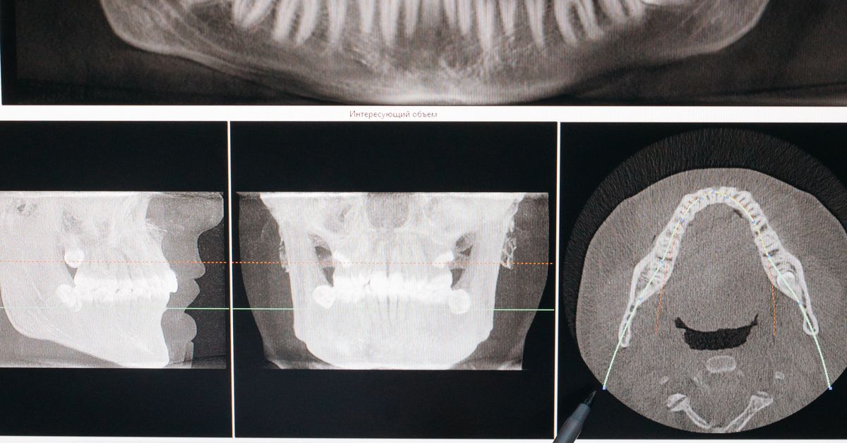
Dental images are not interchangeable. A focused image can show detail around a particular tooth. A panoramic X-ray offers a broad overview of both jaws. A cone beam computed tomography scan, usually called CBCT, gives a three-dimensional view of the area selected for assessment. Your dentist chooses imaging based on the specific question that still needs an answer.
| Record or assessment | What it can help show | Why it may not settle the plan alone |
|---|---|---|
| Focused dental X-ray | Detail near a tooth, its roots, and adjacent bone in the imaged area. | It is a two-dimensional view of a limited region and does not show the full width of the jawbone. |
| Panoramic X-ray | A broad view of the teeth and jaws, useful for an initial overview. | It does not provide the same three-dimensional detail about the implant site’s width or nearby structures. |
| CBCT 3D scan | Three-dimensional relationships, including available bone and the position of structures that may affect implant placement. | It does not replace checking the gums, the bite, the surrounding teeth, or your medical history. |
| Intraoral scan | The surface shape and position of teeth and oral structures for restorative planning. | It records surfaces, not the internal anatomy of the jawbone. |
A patient who arrives with a clear recent CBCT of the appropriate area may have different imaging needs from someone who brings only a panoramic X-ray taken before several teeth were extracted. Even a useful prior scan must be considered in light of changes since it was made. We explain what a dental CBCT scan can contribute in more detail in our imaging guide.
Why a new scan may be recommended: A two-dimensional image may suggest there is room for an implant while leaving the width or shape of the bone uncertain. In the lower jaw, the location of the nerve matters; in the upper back jaw, the relationship to the sinus may matter. A 3D view can help resolve those questions when they are relevant to the proposed site.

How we move from old images to an implant plan
At our Jean-Talon location, the useful first appointment is the one that addresses the decision you actually face. If the question is about a particular missing tooth or an existing implant proposal, a consultation is the relevant appointment type. If several teeth, your gums, or your overall dental condition need a broader assessment, a full exam may be appropriate. Our full exam includes a panoramic X-ray; a consultation for a specific concern may include X-rays or other tests, and complex care may require a full exam.
-
Establish the clinical question
We start with what you want to replace and why. Is the tooth already missing? Is it failing but still in place? Were you told that you might need a graft? Does a denture move when you eat? These are different starting points. The answer guides which records and examinations will be most useful.
-
Review health history and examine the mouth
Your dentist considers medications, relevant health conditions, prior dental treatment, and factors that may affect healing. The examination looks at the gums, the proposed site, neighbouring teeth, available space, and the way the teeth meet. If a natural tooth is still present, whether it can be maintained is part of the decision before replacing it.
-
Assess the existing images
The dentist checks whether each image shows the correct area, provides the detail needed, and still reflects your current situation. A prior X-ray may be helpful for identifying a concern without being sufficient to determine the final position of an implant.
-
Identify only the missing information
If the available images do not answer a necessary anatomical question, additional imaging may be advised. Our Jean-Talon clinic has CBCT 3D imaging and digital scanning capabilities. The dentist can explain why a particular image or scan matters for your case rather than treating more imaging as an automatic next step.
-
Discuss the treatment sequence and final restoration
The plan may involve treating gum disease or infection, removing a failing tooth, assessing bone volume, and deciding when implant placement makes sense. It also needs to address how the implant will support the intended crown, bridge, or denture. For more involved cases, our group practice works with specialists as needed.
This process may answer some questions at the consultation and identify others that require a further examination or image before a definitive plan can be made. A careful assessment is more useful than a premature yes or no based on one picture.
When might other dental care come before an implant?
The images may be adequate, yet the mouth may not be ready for implant treatment. Active gum disease, an unresolved infection, a questionable neighbouring tooth, or limited bone can change the order of care. If the tooth has not been removed, the dentist also needs to assess whether preserving it is a reasonable option.
Gum health and infection
Healthy surrounding tissues matter for an implant that is intended to function over the long term. If you report bleeding, loose teeth, or a history of periodontitis, the gum examination is not a side issue. Addressing active disease may come before replacing teeth. Read more about why gum stability matters before implant treatment.
Bone and the timing of tooth loss
Jawbone can change after tooth loss, so an image taken before an extraction may not represent the site later. If the site appears narrow or close to important anatomy, 3D imaging may help determine whether an implant can be planned there and whether bone reconstruction needs consideration. Being told that you have limited bone does not, by itself, describe every possible approach. It identifies a question that needs site-specific assessment.
Healing and medical factors
Smoking, diabetes, certain medications, and other health factors belong in the discussion because they can influence healing and treatment decisions. Tell the dentist about these before imaging and surgery are planned. Your medical history helps put an X-ray finding into context; the same image can lead to different next steps for different patients.
If you currently have pain, swelling, or a suspected infection: Mention it when booking rather than waiting for a routine implant-planning conversation. The immediate dental problem may need assessment before the replacement tooth can be planned.

Questions worth asking when your X-rays are reviewed
Knowing why a test is proposed helps you understand the treatment plan and compare options. Bring these questions to the visit, particularly if you have already received an implant recommendation elsewhere:
- What can you determine from the images I already have, and what remains uncertain?
- Has anything changed since those images were taken that affects their usefulness?
- If you recommend a CBCT scan, which specific question will it help answer?
- Do the gums, adjacent teeth, and bite need treatment or further assessment first?
- Is the tooth I am considering replacing still a reasonable tooth to maintain?
- Would the planned implant support a crown, bridge, or denture, and how does that affect placement?
- Is bone grafting being considered, and what finding led to that discussion?
- What stages would come before and after implant placement?
- What is included in the estimate, including imaging and the final restoration?
- What other ways could this tooth or these teeth be replaced?
It is especially useful to distinguish an initial opinion from a completed surgical and restorative plan. An early visit can establish a sensible direction, but a decision about implant position should follow the anatomical and restorative assessment needed for that case. If you are comparing recommendations, our article on getting a second opinion before dental implants can help you organize the discussion.
Best practices for making your first implant visit useful
Before the appointment
- Request the actual image files if possible. Include the date of the image and any written interpretation or proposed treatment plan you received.
- Explain the full problem when booking. A single missing molar, a failing front tooth, and an unstable denture may need different appointment scopes.
- Write down what has changed. New swelling, an extraction, new pain, or a tooth that has become loose can affect whether older images still answer the question.
- List medications and health conditions accurately. Implant decisions involve healing as well as anatomy.
During the appointment
- Ask to see the finding. Have the dentist point out what the image shows and where it stops being informative.
- Discuss the finished tooth, not only the implant. Space for a crown, the bite, hygiene access, and the neighbouring teeth matter.
- Clarify the order of care. Ask what needs to happen first, what can wait, and what further information would change the recommendation.
- Keep the options distinct. If a bridge or removable restoration is an option, ask how it differs from an implant for your particular mouth.
The best outcome of a records review is not necessarily an immediate decision to proceed. It is a clear explanation of what is known, what still needs to be checked, and why the next step is appropriate.
Frequently asked questions about existing X-rays and implant planning
Can I bring X-rays from another dentist to the Jean-Talon clinic?
Yes. Bring the images and any related treatment plan to your consultation, or ask the office holding your records about sending the digital files. Tell us when the images were taken and whether the tooth or gums have changed since then. The dentist will assess what the records can contribute to the current decision.
Will I need another panoramic X-ray if I already have one?
Not necessarily. The answer depends on the image’s quality, the area it covers, when it was taken, and what has happened in your mouth since. A panoramic X-ray offers a broad overview, but it may not answer every three-dimensional question involved in implant placement.
Does every implant patient need a CBCT scan?
The need for a CBCT scan is a clinical decision, not something to assume before your appointment. It may be recommended when three-dimensional information about bone or nearby anatomy is needed to plan treatment. Ask your dentist what decision the proposed scan will help make.
If I already have a CBCT scan, can it be used?
Bring it for review if you can. The dentist needs to assess whether it covers the right site, is suitable for the question being asked, and still reflects the site’s current condition. An extraction or other change after the scan may affect how useful it is for the final plan.
Should I book a consultation or a full exam?
A consultation suits a specific implant question or an existing proposal you want to discuss. A full exam may be more suitable when the overall condition of multiple teeth or your gums needs assessment. A full exam at our clinic includes a panoramic X-ray; a consultation may include imaging or tests, and complex care may call for a full exam. Tell us about your existing X-rays when booking so the appointment matches the question.
Can an X-ray alone tell me whether I need bone grafting?
No. An image may raise or clarify questions about bone, but the recommendation also depends on an examination, the proposed implant location, and the intended restoration. A two-dimensional image may not show the bone’s width adequately, which is one reason a dentist may consider 3D imaging.
What if I was told I cannot have an implant because of bone loss?
Bring the images and the explanation you received. The dentist can review which site was assessed, whether more information is needed, and what treatment options make sense in your situation. The question is not simply whether bone loss exists, but how it affects the specific replacement plan.
Can I receive a final implant plan at the first visit?
That depends on how complete and current your records are and what the examination shows. A consultation can clarify likely next steps, but additional imaging, a full exam, or treatment of an existing dental problem may be needed before finalizing the sequence.
Start with the images you have, then decide what is missing
Existing dental X-rays are a useful starting point, not a test of whether you have gathered the perfect records. The right implant assessment connects those images to an examination of the gums, teeth, bone, and bite, then identifies any additional information needed before treatment is planned.
For patients near Jean-Talon, Blanc Dental Center offers implant consultations at its 5001 Rue Jean-Talon O clinic, with CBCT 3D imaging and digital scanning available when appropriate. Bring your images and questions, and we can help you understand the next step without assuming that every previous X-ray must be repeated.
Jean-Talon clinic
5001 Rue Jean-Talon O, Montréal, QC H4P 1W7
(514) 564-4554
Free parking
5001 Rue Jean-Talon O, Montréal, QC H4P 1W7
(514) 564-4554
Free parking
To request an assessment, choose an appointment or call the Jean-Talon clinic and mention that you already have dental X-rays for review.